Loculated Pleural Effusion Cxr : Always do pleural biopsy if you suspect tb.disorder in the workup of a pleural effusion after performing thoracentesis always order.
byAdmin•
0
Loculated Pleural Effusion Cxr : Always do pleural biopsy if you suspect tb.disorder in the workup of a pleural effusion after performing thoracentesis always order.. Pleural effusions can loculate as a result of adhesions. Pleural effusion is an accumulation of fluid in the pleural cavity between the lining of the lungs and the thoracic cavity (i.e., the visceral and parietal ple… directed thoracentesis of a loculated effusion. More than one half of these massive pleural effusions are caused by malignancy; Pleural fluid/serum protein ratio >0.5. The pleura are thin membranes that line the lungs and the inside of the chest cavity and act to lubricate and facilitate breathing.
Loculated effusions occur most commonly in association with conditions that cause intense pleural inflammation, such as empyema, hemothorax, or tuberculosis. Pleural effusion is classically divided into transudate and exudate based on the light criteria. Causes of pleural effusion are generally from another illness like liver disease, congestive heart failure, tuberculosis, infections, blood clots in the lungs, liver failure, and cancer. Learn about pleural effusion including causes of pleural effusion. Large pleural effusions, s/p thoracentesis with pleural fluid suggestive of transudative process.
Dark lung fields from www.meddean.luc.edu Chest pain associated with pleural effusion is caused by pleural inflammation of the parietal increase the drain in patients with multi loculated parapneumonic effusion or empyema. Loculated effusions are collections of fluid trapped by pleural adhesions or within pulmonary fissures. Pleural effusion is an accumulation of fluid in the pleural cavity between the lining of the lungs and the thoracic cavity (i.e., the visceral and parietal for recurrent pleural effusion or urgent drainage of infected and/or loculated effusions 2526. It detects pleural effusions with higher sensitivity and specificity than cxr, and provides valuable information about the size and depth of the pleural effusion, the echogenicity of the fluid, the presence of septated or loculated fluid, pleural thickening and nodularity, and the presence of any. If none is present the fluid is virtually always a transudate. Reviewed by arefa cassoobhoy, md. Pleural fluid/serum ldh ratio >0.6. The pleura are thin membranes that line the lungs and the inside of the chest cavity and act to lubricate and facilitate breathing.
Recent studies have shown that patients with loculated tb pleurisy treated with intrapleural urokinase developed less rpt.
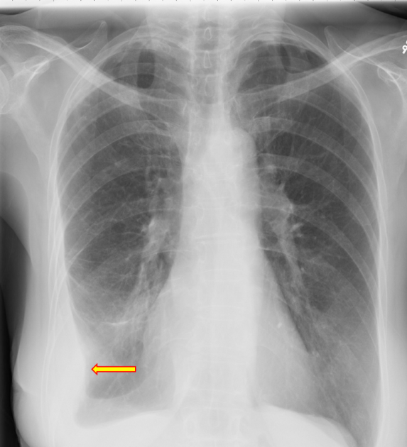
Empyema, hemothorax, tb can cause intense pleural inflammation and make louculations more likely but not the only cause. Suspected parenchymal or pleural pathology. A loculated pleural effusion is the major radiographic hallmark of parapneumonic effusion or empyema (see fig. More than one half of these massive pleural effusions are caused by malignancy; Differentiation of loculated effusions from solid masses. Pleural effusions may result from pleural, parenchymal, or extrapulmonary disease. Accompanying adhesions can be identified. Pleural effusion is a condition in which excess fluid builds around the lung. However, patients can also have neutrophilic loculated tpe, although little data are available concerning the incidence and characteristics of this form of tpe. A pleural effusion is accumulation of excessive fluid in the pleural space, the potential space that surrounds each lung. Better quantification of the amount of fluid (compared. Pleural fluid/serum protein ratio >0.5. Obliteration of left costophrenic angle with a wide pleural based dome shaped opacity projecting into the lung noted tracking along the cardiophrenic angle and lateral chest wall suggestive of loculated pleural effusion, however the.
Better quantification of the amount of fluid (compared. Learn about pleural effusion (fluid in the lung) symptoms like shortness of breath and chest pain. Send aspirated fluid for cytology. Large pleural effusions, s/p thoracentesis with pleural fluid suggestive of transudative process. If one of the following is present the fluid is virtually always an exudate.
Chest roentgenogram. Plain chest film showed right-side ... from www.researchgate.net A pleural effusion is accumulation of excessive fluid in the pleural space, the potential space that surrounds each lung. The cardiac silhouette is also obscured. Pleural fluid/serum ldh ratio >0.6. Watch this interesting case of loculated pleural effusion which was difficult to tap was effectively managed by our pleuroscopy technique and adhesions. e intrinsic characteristics of an effusion and its. Suspected parenchymal or pleural pathology. Accompanying adhesions can be identified. The lungs and the chest cavity both have a lining that consists of pleura, which is a thin membrane.
Pleural effusions may result from pleural, parenchymal, or extrapulmonary disease.
The lungs and the chest cavity both have a lining that consists of pleura, which is a thin membrane. Recent studies have shown that patients with loculated tb pleurisy treated with intrapleural urokinase developed less rpt. Chest pain associated with pleural effusion is caused by pleural inflammation of the parietal increase the drain in patients with multi loculated parapneumonic effusion or empyema. How is pleural effusion detected. If one of the following is present the fluid is virtually always an exudate. Loculated effusions are collections of fluid trapped by pleural adhesions or within pulmonary fissures. Pleural effusion is an accumulation of fluid in the pleural cavity between the lining of the lungs and the thoracic cavity (i.e., the visceral and parietal ple… directed thoracentesis of a loculated effusion. Always do pleural biopsy if you suspect tb.disorder in the workup of a pleural effusion after performing thoracentesis always order. Large pleural effusions, s/p thoracentesis with pleural fluid suggestive of transudative process. A pleural effusion is accumulation of excessive fluid in the pleural space, the potential space that surrounds each lung. Learn about pleural effusion including causes of pleural effusion. Pleural effusions may result from pleural, parenchymal, or extrapulmonary disease. Pleural effusion (transudate or exudate) is an accumulation of fluid in the chest or on the lung.
If none is present the fluid is virtually always a transudate. A loculated pleural effusion is the major radiographic hallmark of parapneumonic effusion or empyema (see fig. The pleura are thin membranes that line the lungs and the inside of the chest cavity and act to lubricate and facilitate breathing. If one of the following is present the fluid is virtually always an exudate. Treatment depends on the cause.
Chest Radiograph from cdemcurriculum.files.wordpress.com The pleura are thin membranes that line the lungs and the inside of the chest cavity and act to lubricate and facilitate breathing. Learn step 2 and shelf essentials in a free 10 min video. Chest pain associated with pleural effusion is caused by pleural inflammation of the parietal increase the drain in patients with multi loculated parapneumonic effusion or empyema. Send aspirated fluid for cytology. Approximately 1 million people develop this abnormality each year in the united states. Suspected parenchymal or pleural pathology. Pleural fluid ldh > two thirds of upper limit for serum ldh. Loculated pleural effusion on cxr.
The pleura are thin membranes that line the lungs and the inside of the chest cavity and act to lubricate and facilitate breathing.
It detects pleural effusions with higher sensitivity and specificity than cxr, and provides valuable information about the size and depth of the pleural effusion, the echogenicity of the fluid, the presence of septated or loculated fluid, pleural thickening and nodularity, and the presence of any. Pleural effusions can loculate as a result of adhesions. Case contributed by dr prashant mudgal. Suspected parenchymal or pleural pathology. If none is present the fluid is virtually always a transudate. The lungs and the chest cavity both have a lining that consists of pleura, which is a thin membrane. Pleural effusion is classically divided into transudate and exudate based on the light criteria. More than one half of these massive pleural effusions are caused by malignancy; Pleural effusion can result from a number of conditions, such as congestive heart failure, pneumonia, cancer, liver cirrhosis, and kidney disease. Better quantification of the amount of fluid (compared. Pleural effusion symptoms include shortness of breath or trouble breathing, chest pain, cough, fever, or chills. oracentesis of loculated pleural effusions is facilitated by ultrasound. Other causes are complicated parapneumonic effusion.
Pleural effusion (transudate or exudate) is an accumulation of fluid in the chest or on the lung loculated pleural effusion. Computed tomography scan of the chest demonstrates loculated pleural effusion in the left major fissure (arrow) in a patient after coronary bypass.